This post is really for the surgeons who treat patellar instability. Every month there is another article describing how to measure MRI or CT scans to determine anatomic factors leading to patellar instability or patellar cartilage overload. It literally becomes alphabet soup with TTTG, C/D index, PT-LTR, etc.
In fact, very few surgeons are going to measure every possible angle on every possible imaging available because it will take too long and it will not necessarily help plan surgery. As one scrolls through an MRI, you get a very good sense of where the patella is tracking and why.
It is then up to the surgeon to determine if they can get away with small surgery, such as an MPFL, or if they need to realign the knee. This can involve a tibial tubercle osteotomy to move the patella medially or distally or a femoral osteotomy to straighten or rotate the knee. The femoral osteotomy has a much higher complication rate and changes the mechanics of the entire lower extremity.
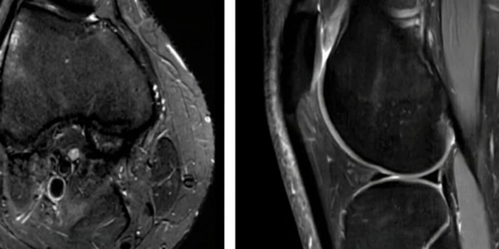
Read the full article in the Orthopaedic Journal of Sports Medicine: Validity of the 2-Image Lateral Trochlear Inclination to Determine Patellar Instability Due to Trochlear Dysplasia.
Image is AI-generated based on an image in the article.