In this Grand Rounds from HSS case report, I shared the story of a 16-year-old female lacrosse player with pain in her left knee. She had no previous traumatic injuries to report but had suffered two noncontact lateral dislocations at ages 11 and 13. To fix her dislocation issues, she had undergone tibial tubercle osteotomy (TTO) combined with a medial patellofemoral ligament (MPFL) reconstruction and particulated cartilage implant to the patella and trochlea.
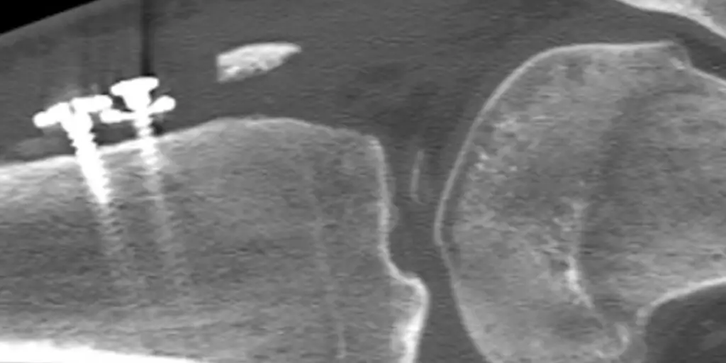
Unfortunately, after the original surgery, the patient reported weakness in her quadriceps with difficulty walking. When I examined her knee, I noted anterior knee crepitus (a grinding noise with a vibration), reduced quadriceps strength, and tenderness over the tibial tubercle. We used computed tomography (CT), magnetic resonance imaging (MRI), and an X-ray of the prior TTO to analyze what was happening. This imaging showed fragmentation of the tibial tubercle shingle and patella alta. I determined that the best approach was arthroscopy and revision TTO. A final X-ray one year after this surgery, we saw complete healing of the tibial tubercle — and our young patient had returned to full range of motion and lacrosse.
Read more about this case report on Grand Rounds from HSS.
(The image is from the case report.)

