We recently conducted a study to identify clinical and radiographic risk factors for medial patellar facet lesions in patients without a history of trauma or patellar instability. Our hypothesis was that a posterior tibial tubercle relative to the trochlear groove would be a risk factor for atraumatic medial patellar facet lesions.
While medial patellar facet lesions have been well-described in the setting of patellar instability, relatively little is known about risk factors for atraumatic medial patellar facet lesions. Read more in the Orthopaedic Journal of Sports Medicine article, “Risk Factors for Atraumatic Medial Patellar Facet Lesions.”
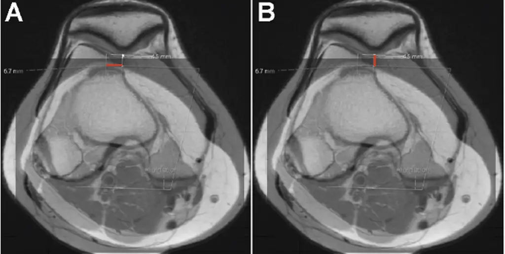
Image from the article: Figure 5. (A) Measuring tibial tubercle-trochlear grove (TTTG) (millimeters) on axial magnetic resonance imaging (MRI) scan. (B) Measuring the tibial tubercle height (in millimeters) on axial MRI scan. Negative values correspond to a tibial tubercle that lies posterior to the trochlear groove.